The Development of a Device for Fluorescent Protein Imaging and Screening
Abstract
Reporter genes are widely used in current animal models in research and analysis. Among the reporter genes, fluorescent proteins are most commonly used due to their availability, stability and diverse color of choice. However, observation and data keeping often become a difficult task due to the pricy equipment, suboptimal photographic quality, and the lack of flexibility for imaging of different fluorescent wavelengths. Not to mention the difficulties in simultaneously observation object with multiple fluorescent wavelengths.
To overcome such obstacles, an imaging device was designed using macro ring light with switchable twin light sources. The macro ring light is compatible with most high-end digital cameras and some internal focusing macro DSLR lenses. To accommodate diverse needs for different fluorescent wavelengths, both light sources can be customized to specific excitation wavelengths. The device is powered by battery/AC duel design to provide flexibility and mobility. For the needs of frequent commute between barriers, the device is also designed to withstand repetitive disinfection.
In addition to the application in fluorescent protein imaging, the device can also be used to detect body fluid in forensic investigation.
Introduction
Fluorescent proteins have many advantageous features such as non-invasiveness, easiness in construct design and detection. Since its first report to use in the research, fluorescent proteins have revolutionized the research in experimentation, data observation and analysis. Tremendous efforts had invested in the improvement of the fluorescent proteins such as their intensity, number of color variant, reduced toxicity which made fluorescent proteins the preferred system to choose for studying gene activities.
Many imaging devices are available for the detection of fluorescent signal. Unfortunately, numerous limitations in the process of fluorescent observation are unavoidable that may consequently influence the data quality. For imaging specific fluorescent signals from a given object, excitation light source and emission light detection device are essential. Furthermore, acceptable imaging outcome often require experience and task, not to mention collecting meaningful data for analysis and standardization for cross laboratories comparisons. Vast majority of the imaging devices for fluorescent detection are either in simplified designed for handheld or bulky structure to ensure required power. Moreover, it can be cumbersome when simultaneous detection of multiple
fluorescent proteins is needed. In addition, most currently available imaging devices rely on human visual judgment for the captured fluorescent signal. Our goal is to overcome obstacles for optical observation. Efforts were made to build an integrated device for easy quality imaging and yet at a fordable price.
Materials and Methods
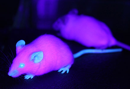
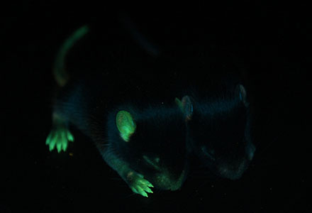
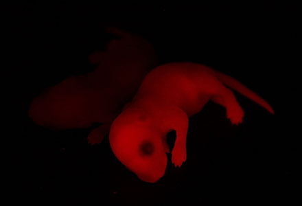
BFP,GFPemd(Emerald) and DsRed are commonly used in research and thus were used in this study. In addition, body fluids such as urine and semen are also included in this study for wider applications in related fields. Key spectrum properties of these fluorescent proteins are listed in (Table 1)
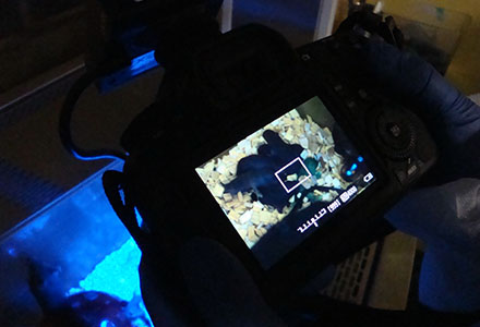
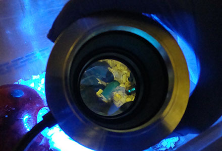
Optical devices: In order to build a flexible light source, we designed a macro ring light which can be mounted directly to the end of DSLR macro lenses, or to the bayonet ring of most high-end compact digital cameras(Figure 1). The macro ring light can be used without camera for direct observation.
The macro ring light also includes the following features: (Figure 2)
- Specific wavelength LED: The ring light has two switchable channels thus up to two wavelengths of excitation light sources can be installed into the same device for convenient observation of multiple fluorescent proteins.
- Light source filter: When peak excitation wavelength is very close to peak emission wavelength, it is necessary to cut off excitation light spectrum to avoid interference of the unwanted fluorescent signals. Often the intensity of excitation light source is much stronger than emission signals.
- Image filter : In order to improve contrast of signal/noise ratio, the purpose of the image filter is to filter out excitation light from emission signals.
Animal models: Several transgenic mouse lines were generated bearing transgene with constitutive promoter expressing specific fluorescent proteins for this study. (Table 1)


